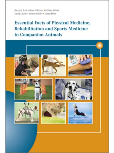
이 책은 총 720페이지, 707장의 사진 그리고 288개의 표로 구성되어 있습니다.
QR code를 이용하여 재생할 수 있는 동영상 168개가 제공됩니다.
오스트리아, 미국, 영국, 독일, 캐나다, 스페인, 브라질, 스위스 등 다양한 국가의 전문가들이 저술하였습니다.
실질적이고 효과적인 매뉴얼과 프로토콜의 제공 뿐 아니라 다양한 상황에서의 주의사항도 다루고 있으며 최신의 기술도 다루고 있습니다.
저자들의 풍부한 경험을 토대로 독자의 빠르고 정확한 발전에 도움이 될 수 있는 책입니다.
목차가 굉장히 상세하고 방대하여 하단에 간단목차와 상세목차를 각각 기재하였습니다.
Main Authors: Barbara Bockstahler, David Levine, Johann Maierl, Darryl Millis, Kathleen Wittek
Contents
1.Foreword 3
1.1The Terms “Physical Medicine” and “Physiotherapy”3
1.2Physical Medicine and Physiotherapy in Veterinary Medicine3
1.3This Book4
2.Physiology and Pathophysiology of Tissues and Healing7
2.1Connective Tissue7
2.1.1Function7
2.1.2Cells7
2.1.3Extracellular Matrix8
Collagen Fibers8
Elastic Fibers and Elastin8
Proteoglycan9
Water9
Non-Collagenous Proteins9
2.1.4Nutrition of the Connective Tissue9
Role of Blood Circulation9
Role of Tissue Loading and Unloading9
2.2Wound Healing10
2.2.1Phases of Wound Healing10
Inflammatory Phase (Day 0-5)10
Reparative Phase (Proliferative, Fibroblastic) (Day 5-21)10
Remodeling Phase (Beginning at Day 21)11
2.3Bones12
2.3.1Function of Bones12
2.3.2Bone Structure12
2.3.3Components13
2.3.4Bone Growth13
2.3.5Importance of Mechanical Stress13
2.3.6Blood Supply13
2.3.7Immobilization and Remobilization14
2.3.8Bone Healing14
Biomechanics14
Primary (Direct) Bone Healing14
Secondary (Indirect) Healing15
Factors Affecting Bone Healing15
2.4Muscles15
2.4.1Structure of Muscles15
2.4.2Types of Muscle Fibres16
2.4.3Blood Supply17
2.4.4Connective Tissue Within the Muscle Belly17
2.4.5Muscle Contraction18
Mechanisms of Muscle Contraction18
Types of Muscle Contractions18
Regulation of Muscle Contractions20
Factors Influencing Muscle Contractions20
Muscle Energy Systems20
Lactate Production During Physical Exertion21
2.4.6Muscle Atrophy21
Disuse Atrophy21
Neurogenic Muscle Atrophy21
Remobilization of Muscles22
2.4.7Muscle Healing22
2.5Tendons22
2.5.1Tendon Structure22
2.5.2Stress-Strain Curve23
2.5.3Myotendinous Junction24
2.5.4Osteotendinous Junction24
2.5.5Immobilization and Remobilization24
2.5.6Tendon Healing24
2.6Ligaments25
2.6.1Ligament Structure25
2.6.2Ligament Healing25
2.7Articular Cartilage25
2.7.1Function and Structure of Articular Cartilage25
2.7.2Nutrition26
2.7.3Role of Stress Stimuli on Cartilage26
2.7.4Cartilage Repair27
2.7.5Immobilization and Remobilization27
2.8Joint Capsule27
2.8.1Response of the Joint Capsule to Disuse/ Immobilization and Remobilization28
2.9Synovial Fluid28
2.10Nervous System28
2.10.1Components of the Nervous System28
2.10.2Central Nervous System28
2.10.3Peripheral Nervous System29
3.Biomechanics - Understand Movement 33
3.1Gait33
3.1.1Basic Principles of the Step Cycle33
3.1.2Normal Dog Gaits33
Walk34
Trot34
Pace35
Gallop35
3.2Joints During Motion36
3.2.1Shoulder Joint36
3.2.2Elbow Joint36
3.2.3Carpal Joint36
3.2.4Hip Joint36
3.2.5Stifle Joint36
3.2.6Tarsal Joint37
3.3Muscles in Motion37
3.3.1Protraction and Retraction38
3.3.2Control of Movement by Flexors and Extensors38
3.3.3Muscles and Gravity38
3.3.4Muscles as Stabilizers39
3.4Vertebral Column in Motion39
3.4.1Cervical Spine39
3.4.2Thoracic Spine39
3.4.3Lumbar Spine39
3.4.4Muscles of the Vertebral Column42
3.5Lameness in Dogs42
3.5.1Forelimbs42
3.5.2Hindlimbs42
3.6Specific Movements43
4.Exercise Physiology 47
4.1Energy Production47
4.1.1Immediate Sources of Energy47
4.1.2Glycolysis47
4.1.3Anaerobic glycolysis47
4.1.4Aerobic glycolysis47
4.1.5Sources of Energy Available While Exercising49
4.1.6The Distribution of Energy49
4.1.7Maximum Oxygen Consumption (VO2 max)49
4.2Muscle Physiology49
4.2.1Types of Muscle Fibers49
4.2.2Types of Muscle Contractions49
4.2.3Musculoskeletal Conditioning49
Endurance Training49
Muscular Strength50
Muscle Fatigue50
4.3Respiration50
4.3.1Anatomy50
4.3.2Control of Ventilation50
4.3.3Mechanisms of Breathing51
4.4Temperature51
Water Balance52
4.5The Cardiovascular System52
4.5.1Blood Pressure and Blood Flow52
4.5.2Function of the Heart52
4.5.3Function of the Arterial Vascular System52
4.5.4Circulatory Changes During Muscular Activity54
4.5.5Changes That Take Place in the Heart ?the Athletic Heart56
4.5.6Exercising and the Cardiovascular System60
4.6Exercise Capacity and Age60
4.7Medical Problems60
4.8Energy Requirement and Nutrition61
5.Pain - Pathophysiology and Management 65
5.1Why is Pain Management so Important in Physiotherapy?65
5.1.1Can Pain be Meaningful?65
5.1.2Possible Stress Reactions Triggered by Pain65
5.2Classifications of Pain65
5.2.1Duration65
5.2.2Origin65
5.2.3Clinical Considerations65
5.3Pain Physiology and Pathophysiology66
5.3.1The Pathway from Triggering Pain to Pain Perception66
5.3.2Segmental Innervation and Function67
Head-Zone67
Mackenzie Zone67
5.3.3Pain Modulation67
Gate Control Theory67
Descending Pain Inhibition67
5.3.4Segmental and Suprasegmental Reflex Responses67
5.3.5Mechanisms Responsible for Prolonged and Increased Pain67
Peripheral Sensitization67
Central Sensitization (Wind-Up)68
Pain Memory68
5.4Measuring Pain, Algesimetry68
5.4.1Algesimetry in Acute Pain68
Numerical Rating Scale (NRS)68
Simple Descriptive Scale (SDS)68
Visual Analog Scale68
Colorado Acute Pain Scale for Dogs and Cats68
Short Form of the Glasgow Composite Measure Pain Scale69
UNESP-Botucatu Multidimensional Composite Pain Scale for Cats69
5.4.2Algesimetry with Chronic Pain69
Helsinki Chronic Pain Index (HCPI)69
Canine Brief Pain Inventory (CBPI)69
Cincinnati Orthopedic Disability Index in Canines (CODI)69
Liverpool Osteoarthritis in Dogs (LOAD)69
Feline Musculoskeletal Pain Index (FMPI)69
Colorado Chronic Pain Scale Dog69
5.5Pain Management70
5.5.1Multimodal Pain Management70
Definition70
Preemptive and Preventative Analgesia70
5.5.2Common Painkillers70
Opioids70
Non-Opioid Analgesics70
Local Anesthetics71
Gabapentin71
Corticosteroids71
5.6Disease Modifying Osteoarthritis Drugs (DMOADs)71
5.7Dietary Supplements71
5.8Other Therapeutical Options71
5.9The Effects of Various Pain Management Procedures72
6.Examination of the Physiotherapy Patient 83
6.1Basics83
6.2Medical History83
6.2.1General Information83
6.2.2History of the Present Problem83
6.3Clinical Examination83
6.4Orthopedic Examination83
6.4.1Posture84
6.4.2Gait84
Degree of Lameness84
Further Assessments84
6.4.3Palpation of the Limbs and Trunk at a Stance84
Specific Tests for Assessment of the Limbs and Back in Stance85
6.4.4Palpation of the Limbs in Recumbency85
Palpation of the Bones and Musculature85
Interphalangeal Joints of Forelimb85
Carpal Joint85
Elbow Joint85
Shoulder Joint85
Interphalangeal Joints of Pelvic Limb86
Tarsal Joint86
Stifle Joint86
Hip Joint86
Lumbosacral Junction87
6.4.5Additional Tests87
General Information87
Range of Motion87
Muscle Mass Measurements91
6.5Neurological Examination93
6.5.1Evaluation at a Stance93
Observation93
Palpation93
6.5.2Gait93
6.5.3Brief Cranial Nerve Examination93
6.5.4Examination of Reflexes93
Pelvic Limb Examination93
Trunk95
Forelimb Examination96
Autonomous Zones97
6.5.5Postural Reflexes97
Hopping97
Hemiwalking97
Wheelbarrowing97
Proprioceptive Positioning Reaction97
Tactile Placing97
6.5.6Interpretation of the Neurologic Examination97
6.6Follow-up Examinations98
6.6.1Guidelines98
6.6.2Specific Things That May Be Assessed Include98
Geriatric Patients98
6.7Cardiovascular Monitoring98
6.7.1Blood Pressure Measurements99
Blood Pressure Measurement Protocol99
Pulse Wave Analysis99
Assessment of Blood Pressure102
Overall Assessment102
6.7.2Clinical Cardiovascular Examination102
6.8Thermography104
6.8.1Basics104
Definition104
Mode of Action104
Sensitivity104
Hair104
Palettes104
6.8.2Thermal Image Interpretation104
Hot Spots105
Cold Spots105
6.8.3Advantages105
6.8.4Limitations105
6.8.5Indications105
6.8.6Contraindications106
6.8.7Videothermography106
6.8.8Patient Management and Imaging Protocol106
7.Passive Range of Motion Exercises and Stretching 109
7.1Basics109
7.1.1Definition109
7.2Passive Range of Motion Exercises (PROM)109
7.2.1Joint Range of Motion (ROM)109
Definition109
Changes in ROM109
7.2.2Muscles Range of Motion (ROM)109
7.2.3Goals109
7.2.4Indications109
7.2.5Contraindications109
7.2.6Clinical Application109
General109
Individual Joints110
Bicycling Motion in Lateral Recumbency112
7.2.7Active Assisted and Active Range of Motion Exercises112
7.3Stretching113
7.3.1Basics113
Definition113
Flexibility113
Contracture113
7.3.2Different Types of Stretching113
7.3.3Goals113
7.3.4Indications113
7.3.5Contraindications113
7.3.6Clinical Application113
Passive Stretching113
Active Stretching114
8.Active Therapeutic Exercises119
8.1A Guide to Using This Chapter119
8.1.1Primary and Secondary Areas of Application119
8.1.2Time Factor119
8.2General Indications119
8.3General Contraindications119
8.3.1Absolute Contraindications119
8.3.2Special Precautions119
8.4Lifting Aids: Moving Straps, Slings and
Harnesses120
8.4.1Applications120
8.4.2Contraindications120
8.4.3Types120
Lifting Harness With Leg Openings120
Chest Harnesses120
Water as a Lifting Aid120
8.4.4Therapeutic Exercises120
8.5Standing With and Without Assistance120
8.5.1Applications120
As a General Rule120
Primary Areas of Application120
Secondary Areas of Application121
Rather Not Recommended for121
8.5.2Contraindications121
8.5.3Time Frame121
8.5.4Biomechanics121
Weight Distribution121
Joints121
8.5.5Therapeutic Exercises121
Exercise 1: Tetraplegic Patients121
Exercise 2: Paraplegic Patients121
Exercise 3: Patients with Decreased Muscle Tone..122
Exercise 4: Patients with Increased Muscle Tone ...122
Exercise 5: Weak Animals122
Additional Exercises122
Frequency and Number of Repetitions for Each Exercise122
8.6Weight Shifting122
8.6.1Applications122
Primary Areas of Application122
Secondary Areas of Application122
Rather Not Recommended for122
8.6.2Contraindications122
8.6.3Time Frame122
8.6.4Biomechanics123
Weight Distribution123
8.6.5Therapeutic Exercises123
Exercise 1: Weight Shifting Using a Gentle
Pushing Technique123
Exercise 2: Weight Shifting Using a Slow Release of Applied Pressure123
Exercise 3: Weight Shifting Using a Quick Release of Applied Pressure123
Exercise 4: Weight Shifting Using Treats124
Exercise 5: Weight Shifting by Lifting One (3-Legged Stance) or Two Limbs124
Exercise 6: Weight Shifting in Water125
Exercise 7: Weight Shifting on a Proprioceptive Wobble Cushion or Trampoline125
Exercise 8: Weight Shifting While Walking126
Frequency and Number of Repetitions for Each Exercise126
8.7Shaking Hands126
8.7.1Applications126
Primary Areas of Application126
Secondary Areas of Application126
Rather Not Recommended for126
8.7.2Contraindications126
8.7.3Time Frame126
8.7.4Biomechanics126
8.7.5Therapeutic Exercises126
Exercise 1: Shaking Hands While Sitting126
Exercise 2: Shaking Hands While Standing126
Exercise 3: Shaking Hands on a Proprioceptive Wobble Cushion126
Frequency and Number of Repetitions for Each Exercise127
8.8Rhythmic Stabilization, Bouncing127
8.8.1Applications127
Primary Areas of Application127
Secondary Areas of Application127
Rather Not Recommended for127
8.8.2Contraindications127
8.8.3Time Frame127
8.8.4Therapeutic Exercises127
Exercise 1: Stable Surface128
Exercise 2: Elastic/Resilient Surface128
Frequency and Number of Repetitions for Each Exercise128
8.9Slow Walks, Walking on a Variety of Surfaces128
8.9.1Applications128
Primary Areas of Application128
Secondary Areas of Application128
Rather Not Recommended for128
8.9.2Contraindications128
8.9.3Time Frame128
8.9.4Biomechanics128
Joints128
8.9.5Therapeutic Exercises128
Exercise 1: Slow Walking128
Exercise 2: Slow Walking with Weight Shifting129
Exercise 3: Walking on a Variety of Surfaces129
Frequency and Number of Repetitions for Each Exercise129
8.10Backwards Walking130
8.10.1Applications130
Primary Areas of Application130
Secondary Areas of Application130
Rather Not Recommended for130
8.10.2Contraindications130
8.10.3Time Frame130
8.10.4Therapeutic Exercises130
Frequency and Number of Repetitions for Each Exercise130
8.11Leg Bands130
8.11.1Applications130
Primary Areas of Application130
Secondary Areas of Application130
Rather Not Recommended for130
8.11.2Contraindications131
8.11.3Time Frame131
8.11.4Types131
8.11.5Therapeutic Exercises131
Frequency and Number of Repetitions for Each Exercise131
8.12Weights Worn on the Back131
8.12.1Applications131
Primary Areas of Application131
Secondary Areas of Application131
Rather Not Recommended for131
8.12.2Contraindications131
8.12.3Time Frame131
8.12.4Biomechanics131
Musculature131
8.12.5Therapeutic Exercises133
Exercise 1: Weights Worn on the Animal’s Back (Standing and Walking)133
Frequency and Number of Repetitions for Each Exercise133
8.13Weights Attached to the Limbs133
8.13.1Applications133
Primary Areas of Application133
Secondary Areas of Application133
Rather Not Recommended for133
8.13.2Contraindications133
8.13.3Time Frame133
8.13.4Biomechanics133
Musculature133
8.13.5Types134
8.13.6Therapeutic Exercises134
Exercise 1: Weights Attached to the Limbs (Standing and Walking)134
Frequency and Number of Repetitions for Each Exercise134
8.14Pulling Exercises134
8.14.1Applications134
Primary Areas of Application134
Secondary Areas of Application134
Rather Not Recommended for134
8.14.2Contraindications134
8.14.3Time Frame135
8.14.4Biomechanics135
8.14.5Types135
8.14.6Therapeutic Exercises135
Exercise 1: Pulling Weight135
Frequency and Number of Repetitions for Each Exercise135
8.15Inclines135
8.15.1Applications135
Primary Areas of Application135
Secondary Areas of Application135
Rather Not Recommended for135
8.15.2Contraindications135
8.15.3Time Frame135
8.15.4Biomechanics135
Joints135
Musculature136
Dogs with Osteoarthritis136
8.15.5Types136
8.15.6Therapeutic Exercises136
Exercise 1: Walking on an Incline, Gentle Slope (<15°/~27%)137
Exercise 2: Walking on an Incline, Steep Slope (>15°/~27%)137
Frequency and Number of Repetitions for Each Exercise137
8.16Declines137
8.16.1Applications137
Primary Areas of Application137
Secondary Areas of Application137
Rather Not Recommended for137
8.16.2Contraindications137
8.16.3Time Frame137
8.16.4Biomechanics138
Joints138
Musculature138
Dogs with Osteoarthritis138
8.16.5Types138
8.16.6Therapeutic Exercises138
Exercise 1: Walking on a Decline, Gentle Slope (<15°/~27%)139
Exercise 2: Walking on a Decline, Steep Slope (>15°/~27%)139
Frequency and Number of Repetitions for Each Exercise139
8.17Ascending Steps139
8.17.1Applications139
Primary Areas of Application139
Secondary Areas of Application139
Rather Not Recommended for139
8.17.2Contraindications139
8.17.3Time Frame139
8.17.4Biomechanics139
Joint139
Musculature140
8.17.5Types140
8.17.6Therapeutic Exercises140
Exercise 1: Ascending Shallow Stairs140
Exercise 2: Ascending Steeper Stairs140
Frequency and Number of Repetitions for Each Exercise 140
8.18Descending Steps140
8.18.1Applications140
As a General Rule140
Primary Areas of Application140
Secondary Area of Application140
Rather Not Recommended for140
8.18.2Contraindications140
8.18.3Time Frame141
8.18.4Biomechanics:141
Joints141
Musculature141
8.18.5Types141
8.18.6Therapeutic Exercises141
Exercise 1: Descending Shallow Steps141
Exercise 2: Descending Steep Steps141
Frequency and Number of Repetitions for Each Exercise 141
8.19Platforms and Steps142
8.19.1Applications142
As a General Rule142
Primary Areas of Application (Depending on the Fundamental Exercise) 142
Secondary Areas of Application (Depending on the Fundamental Exercise)142
Rather Not Recommended for142
8.19.2Contraindications142
8.19.3Time Frame142
8.19.4Types142
8.19.5Therapeutic Exercises142
Exercise 1: Simple Standing Exercises142
Exercise 2: More Strenuous Exercises Performed While Standing142
Exercise 3: Exercises Performed While Walking142
Frequency and Number of Repetitions for Each Exercise142
8.20Walking Across the Slope of a Hill143
8.20.1Applications143
Primary Areas of Application143
Secondary Area of Application143
Rather Not Recommended for143
8.20.2Contraindications143
8.20.3Time Frame143
8.20.4Biomechanics143
8.20.5Therapeutic Exercises143
Frequency and Number of Repetitions for Each Exercise143
8.21Sit-to-Stand144
8.21.1Applications144
As a General Rule144
Primary Areas of Application144
Secondary Area of Application144
Rather Not Recommended for144
8.21.2Contraindications144
8.21.3Time Frame144
8.21.4Biomechanics144
8.21.5Therapeutic Exercise144
Frequency and Number of Repetitions for Each Exercise145
8.22Down-to-Stand145
8.22.1Applications145
Primary Areas of Application145
Secondary Area of Application145
Rather Not Recommended for145
8.22.2Contraindications145
8.22.3Time Frame145
8.22.4Therapeutic Exercises145
Frequency and Number of Repetitions for Each Exercise145
8.23Dancing146
8.23.1Applications146
As a General Rule146
Primary Areas of Application146
Secondary Area of Application146
Rather Not Recommended for146
8.23.2Contraindications146
8.23.3Time Frame146
8.23.4Biomechanics146
Joints146
8.23.5Therapeutic Exercises147
Exercise 1: Dancing Forwards147
Exercise 2: Dancing Backwards147
Frequency and Number of Repetitions for Each Exercise147
8.24Wheelbarrowing147
8.24.1Applications147
As a General Rule147
Primary Areas of Application147
Secondary Area of Application147
Rather Not Recommended for147
8.24.2Contraindications147
8.24.3Time Frame148
8.24.4Biomechanics148
8.24.5Therapeutic Exercises148
Exercise 1: Wheelbarrowing148
Frequency and Number of Repetitions for Each Exercise148
8.25Tunnel, Limbo148
8.25.1Applications148
Primary Areas of Application148
Secondary Area of Application148
Rather Not Recommended for148
8.25.2Contraindications149
8.25.3Time Frame149
8.25.4Biomechanics149
8.25.5Therapeutic Exercises149
Exercise 1: Tunnel, Limbo149
Frequency and Number of Repetitions for Each Exercise149
8.26Balls, Peanuts, Donuts149
8.26.1Applications149
As a General Rule149
Primary Areas of Application149
Secondary Area of Application149
Rather Not Recommended for149
8.26.2Contraindications149
8.26.3Time Frame149
8.26.4Types149
Balls149
Peanuts150
Donuts150
8.26.5Therapeutic Exercises150
Exercise 1: Ball is Centered Under the
Dog’s Body150
Exercise 2: Ball Under the Chest and the Forelimbs150
Exercise 3: Ball Under the Chest and the Hindlimbs151
Exercise 4: Dog Lays on the Ball151
Exercise 5: Dog Stands or Sits on Top of the Ball151
Frequency and Number of Repetitions for Each Exercise152
8.27Wobble Board152
8.27.1Applications152
As a General rule152
Primary Areas of Application152
Secondary Areas of Application152
Rather Not Recommended for152
8.27.2Contraindications152
8.27.3Time Frame152
8.27.4Types152
Multidirectional Board152
Bidirectional Board152
In General152
8.27.5Therapeutic Exercises152
Exercise 1: Back and Forth Movement152
Exercise 2: Right and Left Movement153
Exercise 3: Adding a Proprioceptive
Wobble Cushion153
Exercise 4: 3-Legged Stance153
Exercise 5: Circular Movements153
Rhythm, Amplitude, Speed153
Frequency and Number of Repetitions for Each Exercise154
8.28Proprioceptive Wobble Cushion, Mattress154
8.28.1Applications 154
As a General Rule154
Primary Areas of Application154
Secondary Areas of Application154
Rather Not Recommended for154
8.28.2Contraindications154
8.28.3Time Frame154
8.28.4Types154
Proprioceptive Wobble Cushion154
Mattresses154
8.28.5Functionality154
8.28.6Therapeutic Exercises155
Exercise 1: Standing155
Exercise 2: 3-Legged Stance155
Exercise 3: Standing While Weight Shifting155
Exercise 4: Moving on the Proprioceptive Wobble Cushion and/or Mattress155
Exercise 5: Standing on Two Proprioceptive Cushions156
Exercise 6: Walking over a Proprioceptive Wobble Cushion/Mattress156
Exercise 7: Wobble Board with a Proprioceptive Cushion156
Exercise 8: Sit-to-Stand on a Proprioceptive Wobble Cushion/Mattress156
Exercise 9: Shaking Hands on a Proprioceptive Wobble Cushion/Mattress156
Frequency and Number of Repetitions for Each Exercise156
8.29Trampoline157
8.29.1Applications 157
Primary Areas of Application157
Secondary Area of Application157
Rather Not Recommended for157
8.29.2Contraindications157
8.29.3Time Frame157
8.29.4Types157
8.29.5Therapeutic Exercises157
Exercise 1: Standing on a Moving Trampoline157
Exercise 2: Standing and Weight Shifting Using a Gentle Pushing Technique158
Exercise 3: Standing and Weight Shifting Using a Treat158
Exercise 4: Standing and weight shifting by lifting a limb158
Exercise 5: Rhythmic Stabilization158
Exercise 6: Sit-to-Stand158
Exercise 7: Down-to-Stand158
Frequency and Number of Repetitions for Each Exercise158
8.30Cavaletti Rails159
8.30.1Applications 159
Primary Areas of Application159
Secondary Areas of application159
Rather Not Recommended for159
8.30.2Contraindications159
8.30.3Time Frame159
8.30.4Biomechanics159
Joints159
Muscular Activity159
Dogs with Osteoarthritis159
8.30.5Types160
8.30.6Therapeutic Exercises160
In General160
Exercise 1: Rails Placed on the Ground with a Large Amount of Spacing160
Exercise 2: Rails Placed on the Ground with a Short Amount of Spacing160
Exercise 3: Rails with a Uniform Height160
Exercise 4: Rails with Variable Spacing160
Exercise 5: Rails with Variable Heights160
Additional Exercises161
Frequency and Number of Repetitions for Each Exercise161
8.31Vertical Weave Poles, Circling, Figure-Eight
Walking161
8.31.1Applications161
As a General Rule161
Primary Areas of Application161
Secondary Areas of Application161
Rather Not Recommended for161
8.31.2Contraindications161
8.31.3Time Frame162
8.31.4Type162
8.31.5Therapeutic Exercises162
In General162
Exercise 1: Vertical Weave Poles162
Exercise 2: Circling162
Exercise 3: Figure-Eight Walking162
Frequency and Number of Repetitions for Each Exercise162
8.32Therabands/Elastic Resistance Bands162
8.32.1Applications162
As a General Rule162
Primary Areas of Application162
Secondary Areas of Application162
Rather Not Recommended for162
8.32.2Contraindications162
8.32.3Time Frame162
8.32.4Types163
8.32.5Therapeutic Exercises163
Exercise 1: Targeting Specific Muscle Groups163
Exercise 2: Assisting with Movement163
Frequency and Number of Repetitions for Each Exercise163
8.33Clinical Applications of Therapeutic Exercises ...163
8.33.1ROM and Joint Function163
8.33.2Limb Loading163
8.33.3Proprioception and Balance164
8.33.4Musculature ? Activating, Preserving, Building ...164
9.Land Treadmill167
9.1Types of Treadmills167
9.1.1Normal Treadmill167
9.1.2Treadmills with a Hoist and Harness System (Sling Lifting Device)167
9.2Applications 168
Primary Applications168
Secondary Applications168
Rather Not Suitable for168
9.2.1Contraindications168
9.3Standing Exercises168
9.3.1Applications 168
General168
Primary Applications168
Secondary Applications168
Rather Not Suitable for168
9.3.2Contraindications168
9.3.3Time Frame168
9.3.4Therapeutic Exercises169
Exercise 1: Weight Shifting Incorporating Gentle Nudges169
Exercise 2: Weight Shifting Using Slow Release of Applied Pressure169
9.4.8Therapeutic Exercises to Improve Strength and Conditioning171
General171
Exercise 1: Changing the Speed172
Exercise 2: Increasing the Time172
Exercise 3: Inclined or Declined Slope172
Exercise 4: Weights Attached to the Limbs172
Exercise 5: Weights Worn on the Back172
Exercise 6: Resistance Bands172
Exercise 7: Stepping Over an Outstretched Hand... 172
Exercise 8: Walking on the Rear Limbs172
Exercise 9: The Combination of Load, Speed and Time172
Cooling Down172
10.Aquatic Therapy175
10.1Basics175
10.1.1Relative Density175
10.1.2Buoyancy175
10.1.3Hydrostatic Pressure175
10.1.4Viscosity and Resistance175
10.1.5Surface Tension175
10.1.6Technical Safety175
10.2Indications176
10.3Contraindications176
10.4Importance of Water Temperature176
10.4.1Underwater Treadmill176
10.4.2Swimming176
10.5Water Depth and Speed of Movement176
10.5.1Human176
10.5.2Horses176
10.6VO2, Heart Rate, Energy Consumption177
10.6.1Underwater Treadmill177
10.6.2Swimming177
10.7Musculature178
10.7.1Underwater Treadmill178
Human178
Horses178
10.7.2Swimming178
10.8Limb Loading, Stride Parameters178
10.9Kinematics178
10.9.1Underwater Treadmill178
10.9.2Swimming178
10.10Balance179
10.11Osteoarthritis180
10.11.1Humans180
10.11.2Horses180
10.11.3Dogs180
10.12Overweight180
10.12.1Humans180
10.12.2Dogs180
10.13Underwater Treadmill ? Standing Exercises180
10.13.1Applications180
As a General Rule180
Primary Areas of Application180
Secondary Areas of Application180
Rather Not Appropriate for180
Examples Include180
10.13.2Contraindications180
10.13.3Time Frame181
10.13.4Therapeutic Exercises181
Exercise 1: Standing in Water181
Exercise 2: Weight Shifting Using the Motion of Water181
Exercise 3: Weight Shifting Using Slow Release of Applied Pressure181
Exercise 4: Weight Shifting Using a Quick Release of Applied Pressure181
Exercise 5: Weight Shifting Using Treats181
Exercise 6: Weight Shifting by Lifting One (3-Legged Stance) or Two Limbs182
Exercise 7: Bicycling While Standing182
Exercise 8: Inclines182
Frequency and Number of Repetitions for Each Exercise183
10.14Underwater Treadmill ? Walking Exercises183
10.14.1Applications183
As a General Rule183
Primary Areas of Application183
Secondary Areas of Application183
Rather Not Appropriate for183
10.14.2Contraindications183
10.14.3Time Frame183
10.14.4Therapeutic Exercises183
Exercise 1: Assisted Walking183
Exercise 2: Walking on the Underwater Treadmill...183
Exercise 3: Weight Shifting While Walking184
Exercise 4: Apply Pressure to Specific Muscle Groups184
Exercise 5: Changing the Tempo184
Exercise 6: Exaggerate the Stride185
Exercise 7: Stepping Over an Outstretched Hand...185
Exercise 8: Varying Water Depths185
Exercise 9: Increasing the Duration185
Exercise 10: Walking on an Incline185
Exercise 11: Weights Attached to the Limbs185
Exercise 12: Weights Attached to the Back185
Exercise 13: Apply Physical Resistance During Active Exercises185
Exercise 14: Therabands185
Exercise 15: Working with Countercurrents186
Exercise 16: Combining Various Loads, Speed and Time186
Frequency and Number of Repetitions for Each Exercise186
10.14.5Post Exercise186
10.15Exercising in a Pool186
10.16Therapeutic Floating Exercises186
10.16.1Applications186
Primary Areas of Applications186
Secondary Areas of Application186
Rather Not Appropriate for186
10.16.2Contraindications186
10.16.3Time Frame187
10.16.4Therapeutic Exercises187
Exercise 1: Floating187
Exercise 2: Passive Exercises for Dogs Floating in Water187
Exercise 3: Carefully Position the Floating Dog Slightly off Balanced in the Water187
Exercise 4: Apply Pressure to the Foot Pads187
10.17Therapeutic Exercises While Swimming in a Pool187
10.17.1Applications187
Primary Areas of Application187
Secondary Areas of Application187
Rather Not Appropriate for187
10.17.2Contraindications187
10.17.3Time Frame187
Exercise 1: Swimming187
Exercise 2: Swimming with Weights187
Exercise 3: Swimming Against Resistance187
Exercise 4: Swimming Against a Countercurrent187
10.18Additional Exercises in a Pool188
10.18.1Applications188
Primary Areas of Application188
Secondary Areas of Application188
Rather Not Appropriate for188
10.18.2Contraindications188
10.18.3Time Frame188
Exercise 1: Balancing on a Kickboard188
11.Joint Mobilization in Canines191
11.1Basics191
11.1.1Definition of Manual Therapy191
11.1.2Basic Principles191
Types of Motion191
Concave and Convex Joint Relationships191
Joint End Feels191
11.2Techniques192
11.3Indications192
11.4Contraindications and Precautions192
11.5Clinical Application192
11.5.1Shoulder Joint193
11.5.2Elbow Joint193
11.5.3Carpus194
11.5.4Hip Joint196
11.5.5Stifle Joint197
11.5.6Tarsus197
11.5.7Spine198
12.Massage Therapy203
12.1Basic203
12.1.1Definition203
12.1.2Reasons for Massage203
12.1.3Biological Effects203
Hyperemia203
Mechanical Effects203
Endogenous Endorphins203
Neurological Effects203
12.1.4Benefits of Massage203
12.1.5Indications203
12.1.6Contraindications and Precautions203
12.2Massage Techniques204
12.2.1Effleurage (Gliding, Stroking)204
Effects204
Procedure204
12.2.2Petrissage (Kneading)204
Effects204
Procedure204
12.2.3Rubbing (Friction)205
Effects205
Procedure205
Circular Pressure (a Subset of Friction Massage).. 205
12.2.4Shaking, Vibration205
Effects205
Procedure205
12.2.5Percussion/Tapotement (Clapping)206
Effects206
Procedure206
12.2.6Massage Variables206
Hands206
Pressure206
Duration206
Frequency206
Shape206
Speed206
Direction206
12.3Use of Instruments in Massage206
12.3.1Biological Effects206
Tissues206
Motor Control206
12.3.2Safety When Using Instruments206
12.3.3Treatment Variables207
Tool Grip/Handling207
Duration207
Frequency207
Depth207
Vectors207
Fluid Capture207
12.3.4Indications and Contraindications207
12.4Conducting a Massage Session208
12.4.1Environment208
12.4.2Procedure208
12.5Recovery208
12.6Athletic Conditioning208
12.6.1Pre-Event208
12.6.2Post-Event208
12.7Myofascial Trigger Points209
12.7.1Definition209
12.7.2Pathogenesis209
12.7.3Diagnosis209
12.7.4Treatment209
Non-Invasive209
Invasive209
Treatment Frequency210
12.7.5Contraindications and Precautions210
12.8Traditional Chinese Veterinary Medicine (Tuina and Acupressure)
12.8.1Tuina210
12.8.2Acupressure210
13.Superficial Thermal Modalities213
13.1Definition213
13.2Superficial Heat213
13.2.1Definition213
13.2.2Physics of Thermotherapy213
Hot Packs213
Infrared Heat Lamps213
Therapeutic Ultrasound213
Additional Methods213
13.2.3Biological Effects213
Blood Vessels213
Connective Tissues213
Musculature213
Nerves213
Pain213
13.2.4Indications213
13.2.5Contraindications and Precautions213
13.2.6Equipment214
Hot Packs214
Infrared Heat Lamps214
Additional Methods215
13.2.7Treatment Time and Frequency215
13.3Cryotherapy215
13.3.1Definition215
13.3.2Biological Effects215
Vasoconstriction215
Cell Metabolism215
Hunting Response215
Pain Relief215
Reduction in Muscle Spasms215
13.3.3Indications215
13.3.4Contraindications and Precautions215
13.3.5Equipment215
Ice Packs215
Cold Packs215
13.3.6Treatment Time and Frequency216
14.Therapeutic Ultrasound219
14.1Basics219
14.1.1Definition219
14.1.2Ultrasound and Medium219
Ultrasound and Interfaces219
Absorption219
14.1.3Generating Ultrasound Waves219
14.2Sound Parameters219
14.2.1Intensity219
14.2.2Ultrasound Modalities219
14.2.3Pulse Ratio and Duty cycle220
14.2.4Mode and Intensities220
14.2.5Frequency220
14.3Biological Effects220
14.3.1Thermal Effects220
14.3.2
14.4Mechanical Effects220
Indications221
14.5Contraindications and Precautions221
14.6Clinical Application221
14.6.1Size of the Treatment Area221
14.6.2Frequency221
14.6.3Mode221
14.6.4Intensity221
14.6.5Treatment Time221
14.6.6Treatment Frequency and Time221
14.6.7Coupling222
14.6.8Application Techniques222
14.7Phonophoresis223
14.7.1Definition223
14.7.2Biological Basis223
14.7.3Indications223
14.7.4Contraindications223
14.7.5Clinical Application223
15.Electrical Stimulation227
15.1Basics227
15.1.1Definition227
15.1.2Terminology227
15.1.3Electrical Current Parameters227
Pulse Parameters227
Pulse and Phase Duration227
Waveform227
Pulse Interval (ON/OFF Time)227
Ramp Up/Down228
Specific Types of Current228
15.2Biological Effects228
15.2.1Motor Response228
15.2.2Muscle228
15.2.3Blood Flow228
15.2.4Analgesia228
15.3Indications229
15.4Contraindications and Precautions229
15.5Electrodes229
15.6Pain Therapy229
15.6.1TENS229
High-Frequency, Low-Intensity TENS229
High-Intensity, Low-Frequency TENS229
Burst TENS230
Modulated TENS230
15.6.2Interferential and Pre-Modulated Interferential Currents230
15.6.3Electrode Placement230
15.6.4Treatment Parameters230
Choosing the Appropriate Type of Electrical Stimulator230
Planning the Treatment Frequency, Intensity and Duration230
15.6.5Select the Treatment Area231
Local Placement of the Electrode231
Segmental Placement of the Electrodes232
15.6.6Treatment Procedure233
Clinical or Home Treatment?234
15.7Muscle Reeducation234
15.7.1Clinical Use234
15.7.2Contraindications and Precautions234
15.7.3Voluntary Verses Electrically Provoked Contractions234
15.7.4A Summary of Stimulator Parameters235
Duration235
Frequency235
Pulse Interval (ON/OFF Time)235
Amplitude235
Ramp235
Placement of the Electrodes235
Factors That Influence Contractions235
15.7.5General Recommendations236
15.7.6Side Effects/Adverse Effects236
15.8Electrotherapy in Patients with Flaccid Paralysis236
15.9Iontophoresis236
16.Extracorporeal Shock Wave Therapy (ESWT) 239
16.1Definition239
16.2Technical Aspects239
16.2.1Shockwave Generation239
Focused239
Radial239
16.3Biological Effects239
16.4Indications240
16.5Contraindications and Precautions240
16.6Treatment Procedure241
16.6.1Basics241
16.6.2Patient Preparation241
16.6.3Treatment241
16.6.4Sample Focused Shockwave Protocols241
16.6.5Sample Radial Shockwave Protocols241
16.6.6Treatment Frequency241
16.6.7Side Effects of ESWT241
17.Laser Therapy245
17.1Basics245
17.2Laser Emission and Tissues245
17.3General Information About Laser Emission
and Treatment Parameters247
17.3.1Pulsed vs. Continuous lasers247
17.3.2Treatment Parameter Considerations248
Wavelength248
Energy Density248
Pulsed Laser248
Application Technique248
Depth of Penetration248
17.3.3Recommendations248
17.4Biological Effects248
17.4.1Photochemical Interaction249
17.4.2Photothermal Interaction249
17.4.3Photomechanical Interaction249
17.5Therapeutic Effects250
17.5.1Anti-Inflammatory Effect250
17.5.2 Anti-Edema Effect ............ 250
17.5.3 Analgesic Effect ........... 250
17.5.4 Stimulation of Tissue Repair .......... 250
17.6 Clinical Use of Laser Therapy .........251
17.6.1 Prior to Use ................251
17.6.2 Treatment ..............251
17.6.3 Suggested Doses ..............251
17.6.4 Precautions .............. 252
18. Nuclear Magnetic Resonance Therapy 255
18.1 Basic Information ............ 255
18.2 Magnetic Resonance Therapy ........ 256
18.3 Scientific Principles ............. 256
18.3.1 Clinical Studies ............ 258
Back Pain ............... 258
Osteoarthritis ............ 258
Osteoporosis ............. 258
18.3.2 In Vitro Studies ........... 258
Fibroblasts ................. 258
Chondrocytes ............ 258
Chronobiology ............... 258
Research in Progress ............ 258
18.4 Indications ................ 258
18.5 Contraindications and Precautions ....... 258
18.6 Clinical Applications ............ 259
19. Kinesiology Taping 263
19.1 Definition .............. 263
19.2 Principles of Kinesiology Taping ........ 263
19.2.1 What is Tape? ................ 263
How is Tape Made? ........... 263
19.2.2 Biological Effects of Kinesiology Taping ....... 263
Tissue Decompression ......... 263
Stimulation of Sensory Nerves ........ 263
Nociception ............... 264
19.2.3 Indications ............... 264
19.2.4 Contraindications/Precautions ........ 264
19.3 How to Tape Effectively .......... 264
19.4 Taping Techniques ........... 264
19.4.1 Taping for Pain Mitigation ........... 265
Goal ............... 265
Procedure ............. 265
Indications ................. 265
19.4.2 Taping for Inflammation/Edema ....... 266
Goal ............... 266
Procedure .............. 266
Indications ................. 266
19.4.3 Taping for Neurosensory Awareness/Posture ..... 267
Goal ............... 267
Procedure .............. 267
Indications ................. 267
19.4.4 Once the Tape is Applied ............ 267
20. Veterinary Orthotics and Prosthetics in Practice 271
20.1 Basics .................271
20.1.1 Definitions .............271
Orthotics .................271
Prosthetics .............271
20.2 Orthoses ................271
20.2.1 Types of Orthoses ............ 272
20.2.2 Clinical Case Scenarios .......... 272
Rehabilitative and Prophylactic orthosis ..... 272
Functional orthosis ........... 272
Prophylactic ............... 272
20.2.3 Casting for Custom Rigid Orthoses ....... 273
Materials for Casting ............. 273
Preparing for Casting .............274
Patient Preparation for Casting ........274
Patient Positioning .............274
Casting for an Orthosis ............. 275
Limb Positioning During Casting .......... 275
Final Preparation of Cast Mold .........276
20.2.4 Fitting and Introducing an Orthosis ...... 277
Initial Orthotic Fitting .......... 277
Initial Orthotic Wearing Instructions ...... 277
20.2.5 General Orthotic Wear ............ 277
20.2.6 Possible Orthotic Complications ....... 278
Risk Factors ............... 278
Prevention ............. 278
Diagnosis for Mechanical Improper Alignment .. 278
Correction of the Device .......... 279
20.3 Prosthesis ................ 280
20.3.1 Prosthesis Case Selection .......... 280
20.3.2 Prosthesis Manufacturers and Device Types .. 280
20.3.3 Exoskeletal and Endoskeletal Prostheses .... 280
20.3.4 General Types of Prosthesis ........... 280
20.3.5 Case Selection and Surgical Planning for Prostheses ................ 280
20.3.6 Casting and Measuring for a Prosthesis ...281
Preparation .................281
Submitting the Final Cast Mold ........281
20.3.7 Fitting and Introduction ...........281
General Tips for Prosthesis Wear ....... 282
Introducing the Prosthesis ........... 282
Monitoring the Prosthesis and Residuum ...... 282
Prosthesis Modifications .......... 283
20.4 Therapeutic Exercise with Orthotics and Prosthetics ............... 283
21. Regenerative Medicine and Biological Treatment Approaches 287
21.1 Why Regenerative and Biological Therapies? .. 287
21.2 Platelet Rich Plasma (PRP) ......... 287
21.2.1 Basics ................ 287
21.2.2 Production of PRP ........... 287
21.2.3 Uses for PRP ............ 288
21.2.4 Clinical Application of PRP ......... 288
21.3 IRAP ............... 289
21.4 Stem Cells 289
21.4.1 Production 289
In-House Systems 289
Culture Expansion 289
21.4.2 Legal Regulations 290
USA 290
Europe/Asia 290
21.4.3 AD-MSC (SVF) 290
Production 290
21.4.4 BM-MSC (BMAC) 290
Production 290
21.4.5 Advantages and Disadvantages 290
AD-MSC (SVF) 290
BM-MSC (BMAC) 291
21.4.6 Uses for MSC 291
21.4.7 Clinical Application of MSC 291
21.4.8 Combined Stem Cells and PRP Injections 291
21.5 Principles of Joint Injections 292
21.5.1 Basics 292
21.5.2 Injecting Specific Joints 292
Shoulder 292
Elbow 292
Carpus 293
Hip 293
Stifle 293
Tarsus 294
21.6 Research in PRP and MSC 294
21.7 Rehabilitation Following Treatment with Regenerative Therapies 297
21.7.1 Principles 297
21.7.2 Special Considerations 297
Laser 297
Cryotherapy 297
Other Modalities 297
Sample Rehabilitation Protocol 297
22. Magnet Therapy - Static Magnetic Fields (SMF) and Pulsed Electromagnetic Fields (PEMF)301
22.1Introduction301
22.2Basics301
22.3Types of Magnetic Fields301
22.3.1Static or Permanent Magnetic Fields (SMF/PMF)301
Research to the Effects of SMF302
22.3.2Pulsed Electromagnetic Fields (PEMF)302
Research to the Effects of PEMF302
22.4Method of Application304
22.5Contraindications305
23. Diet and Weight Management in Physiotherapy 309
23.1Obesity in the Rehabilitation Patient309
23.1.1Biology of Obesity309
Hormones and Appetite309
Adipose Tissue309
Endocrinology of Adipose Tissue309
23.1.2Common Weight Loss Protocols310
Body Condition Score310
Diet History310
Calculation for Weight Loss311
Expected Weight Loss311
Once Ideal Weight is Achieved311
23.1.3Role of Exercise in Weight Loss and
Osteoarthritis312
Exercise and Joints312
23.1.4Dietary Approaches for Success312
Prescription Diets312
Drugs That Block Fat Absorption312
Over the Counter Foods Versus Therapeutic Diets. 312
Reduced Fat313
Protein and Lean Mass313
Fiber and Extrusion314
Adjustments to the Current Feeding Regimen314
Water and Canned Food314
Cooked/Raw Diet (BARF)314
Nutraceuticals and Obesity314
Treats314
23.2Nutrition in Gerontology and Rehabilitation315
23.2.1Sarcopenia315
Protein Requirement in Aged Dogs315
Exercise and Sarcopenia315
23.2.2Nutraceutical Use in Canine Osteoarthritis316
Glucosamine and Chondroitin Sulfate316
Green Lipped Mussel316
Undenatured Collagen-II316
Avocado/Soybean Unsaponifiables317
Omega-3 Fatty Acids317
Boswellia Serrata318
Turmeric Root318
Dimethylsulfone/Methylsulfonylmethane (MSM)318
Collagen Hydrolysate318
African Devil’s Claw318
Elk Antler Velvet318
23.2.3Dietetic Foods and Osteoarthritis319
23.3Basic Nutrition for the Sporting Dog319
23.3.1Body Condition Score in Sporting Dogs319
23.3.2Energy and Activity319
Fuels for ATP Generation: Fat and Carbohydrate319
Protein320
Fat and Carbohydrates320
Electrolytes, Minerals and Vitamins320
Vitamins320
24. The Home Environment 325
24.1 In the Home .............. 325
24.1.1 Nonslip Flooring .............. 325
24.1.2 Stairs ................. 325
24.1.3 Resting and Sleeping Area ......... 325
24.1.4 Bed and Sofa ............ 325
24.1.5 Feeding Area ................ 325
24.2 The Car .................. 325
24.3 Walking Aids ............ 325
24.4 Abraded Nails and Dorsal Paws ......... 326
25. The Owner’s Role in Physiotherapy 329
25.1 Basic Concept .............. 329
25.2 Prerequisites ............ 329
25.2.1 Owner ................ 329
25.2.2 Animal ............... 329
25.2.3 Therapist ............... 329
25.3 Home Treatment Modalities ........... 329
25.3.1 Superficial Thermotherapy ......... 329
25.3.2 Massage ................ 329
25.3.3 Therapeutic Exercises ............ 329
25.3.4 Electrical Stimulation ......... 330
25.4 Sample Home Treatment Protocols ...... 330
25.4.1 Arthritis and Spondylosis .......... 330
25.4.2 Cranial Cruciate Rupture
(After Suture Removal) ............ 330
25.4.3 Intervertebral Disk Surgery (After Suture Removal) ..... 330
25.4.4 Degenerative Myelopathy ............331
25.4.5 Fractures ................331
26. Choosing the Right Therapy - Practical Tips from the Expert 335
26.1 Orthopedic Conditions, Surgical ....... 335
26.1.1 Acute, Early Stages - Typically Day 0-10 ... 335
26.1.2 Middle Stages - Typically Day 10-45 ..... 335
26.1.3 Late Stages ............... 335
26.2 Orthopedic Disorders, Non-Surgical ..... 336
26.2.1 Acute Stages- Typically Day 0-10 ....... 336
26.2.2 Middle Stages - Typically Day 10-45 .... 336
26.2.3 Late Stages .......... 337
26.3 Neurological Disorders, Surgical ....... 337
26.3.1 Acute Stages - Typically Day 0-21 ..... 337
26.3.2 Middle Stage - Typically Day 7-45 ......... 338
26.3.3 Late Stages .......... 338
26.4 Neurologic Conditions, Non-Surgical ...... 338
26.4.1 Neurologic Rehabilitation of Non-Surgical Severe Deficits ..... 339
26.4.2 Neurologic Rehabilitation of Non-Surgical Moderate Deficits .... 339
26.4.3 Neurologic Rehabilitation of Non-Surgical Mild Deficits ..... 339
27. The Athletic Dog in Physiotherapy 343
27.1 Types of Canine Sports ........... 343
27.1.1 Agility ................ 343
Common Injuries ...... 343
27.1.2 Hoopers Agility ............ 344
27.1.3 Mobility .................. 344
27.1.4 Obedience ................. 344
Common Injuries ........ 344
Working Trials ...... 344
Rally Obedience ........ 344
27.1.5 Dog Dancing ........ 344
27.1.6 Lunging ........... 344
27.1.7 Tracking ......... 344
27.1.8 Mantrailing ........ 344
27.1.9 Search and Rescue Dogs ............ 345
Common Injuries .......... 345
27.1.10 Hunting Dogs ........ 345
Common Injuries ....... 345
27.1.11 Flyball .......... 345
Common Injuries .......... 345
27.1.12 Frisbee Dog/Disc Dog ...... 345
Common Injuries ........ 346
27.1.13 Schutzhund/IPO Sport ........... 346
27.1.14 Canine Herding ............ 346
27.1.15 Treibball ............... 346
27.1.16 Turnierhundsport (THS) .......... 346
27.1.17 Drafting ................. 346
27.1.18 Canicross .............. 346
27.1.19 Bikejoering, Scooterjoering/Scootering, Skijoering, Trike-, Wagon- or Cart Racing ..... 346
27.1.20 Sled Dogs (Mushing) .......... 347
Common Injuries ..... 347
27.1.21 Greyhound Racing ......... 347
Common Injuries ....... 347
27.2 Specific Injuries in Canine Athletes ...... 348
27.2.1 Injury to the Muscles ? General Information .. 348
Partial or Complete Muscle Tear ........ 348
“Muscle Strains” ......... 349
Muscle Contusion .......... 350
27.2.2 Injuries of Specific Muscles ........ 350
Insertional Tendinopathy of the Supraspinous ... 350
Contracture of the Infraspinosus Muscle ... 350
Tendinopathy of the Proximal Biceps Tendon .....351
Biceps Brachii Avulsion .......351
Luxation of the Biceps Tendon ....... 352
Medial Humeral Epicondylitis ..... 352
Insertional Tendinopathy of the Flexor Carpi Ulnaris Muscle ..... 353
Avulsion of the Long Head of the Triceps Brachii Muscle ... 353
Injury of the Iliopsoas Muscle ....... 353
Injury to the Adductor Longus Muscle .... 354
Tear of the Gracilis Muscle ....... 354
Myopathy of the Semitendinosus and Semimembranosus Muscle .. 355
Injury of the Long Digital Extensor Muscle ..... 355
Tear/Avulsion of the Gastrocnemius Muscle ...... 356
Gastrocnemius Tendinopathy .... 356
Tear or Rupture of the Common Calcaneal Tendon (Achilles Tendon) .... 357
Achillodynia ........ 357
Superficial Digital Flexor Displacement ...... 357
27.2.3 Injuries to the Ligaments ........... 358
Torn Ligaments ...... 358
Sprained Ligaments .... 358
27.2.4 Fractures 359
Carpal Fractures 359
Tarsal Fractures 359
Tarsal/Hock Injuries 360
Nonarticular Fractures of the Long Bones 360
Stress Fractures 361
27.2.5 Shoulder 361
Medial Shoulder Syndrome and Medial Shoulder Instability 361
27.2.6 Elbow 362
Medial Elbow Compartment Injuries 362
27.2.7 Carpal Joint 363
Carpal Joint Sprain 363
Carpal Hyperextension 363
27.2.8 Stifle 363
Traumatic Cranial Cruciate Ligament Rupture 363
Posterior Cruciate Rupture, Caudal Cruciate Ligament Tear 364
27.2.9 Tarsus 365
Plantar Ligament Rupture 365
27.2.10 Injuries to the Paw 365
27.2.11 Injuries to the Digits 365
Phalangeal Luxations with Sprain or Tear of the Collateral Ligaments 366
Superficial Digital Flexor Tendon Ruptures
(“Dropped Toe, Flat Toe”) 366
Deep Digital Flexor Tendon Ruptures
(“Knocked up, Kicked up Toe”) 366
Tendinitis of the Digital Flexors 366
27.2.12 Acute Caudal Myopathy 366
27.2.13 Acute Hindlimb Paralysis 366
27.3Path from Rehabilitation Back to Training367
27.3.1Advanced Exercises to Complement Those Described in the Chapter Entitled “Active Therapeutic Exercises”367
Shake Hands367
Standing Uphill/Downhill368
Slow Walks/Walking on Various Surfaces368
Walking Backwards368
Weights368
Pulling368
Walking Uphill368
Walking Downhill368
Stairs368
Slopes - Walking Perpendicular to the Slope368
Sit-to-Stand368
Down-to-Stand369
Wheelbarrow and Dancing369
Balls/Peanut Balls/Donuts369
Mats/Proprioception Cushion/Trampoline369
Weaves/Circling/Figure-8s369
Therabands370
27.3.2Water Therapy370
Underwater Treadmill370
Swimming370
27.3.3Land Treadmill370
27.3.4Additional Exercise Options370
Tugging370
Digging370
Beg-to-Standing-on-Hind-Legs370
Sit-Ups370
Side Sit-Ups370
Squats371
Recall371
27.3.5Training Using Various Agility Obstacles371
Tunnels and Pipe Tunnels371
A-Frame371
Dog Walk371
Seesaw371
Jumping Obstacles371
Weave Poles372
27.3.6Warming up and Cooling down372
Warming up372
Cool Down372
28.Treatment Plans375
28.1A Guide to Using This Chapter375
28.1.1Introduction375
28.1.2The Path Leading to a Treatment Plan375
Step 1: Create a Problem List375
Step 2: Choose a Modality375
Step 3: Choosing Therapeutic Exercises376
Step 4: Distinct Features of Specific Diseases377
28.2General Treatment Plan for the Forelimb378
28.2.1Pain378
28.2.2Swelling378
28.2.3Tissue Healing and Regeneration378
28.2.4Secondary Muscular Tension379
28.2.5Range of Motion and Joint Function379
28.2.6Limb Loading380
28.2.7Proprioception and Balance381
28.2.8Musculature: Activation of Atrophic Musculature and Promotion of Strength and Endurance383
28.3Scapula384
28.3.1Anatomy384
Bone Type384
Bones384
Joints384
Palpable Bone Structures384
Movement384
Muscles384
28.3.2Fractures389
Definition389
Clinical Signs389
Diagnosis389
Conservative Treatment389
Surgical Treatment389
Special Considerations Within the Rehabilitation Plan389
28.3.3Luxation390
Definition390
Clinical Signs390
Diagnosis390
Conservative Treatment390
Surgical Treatment390
Special Considerations Within the Rehabilitation Plan390
28.3.4Avulsion of the Long Head of the Triceps
Brachii Muscle390
Definition390
Clinical Signs390
Diagnosis390
Conservative Treatment390
Surgical Treatment391
Special Considerations Within the Rehabilitation Plan391
28.4 Shoulder Joint .....391
28.4.1 Anatomy ...391
Joint Type .......391
Bones .....391
Articulation ...... 392
Joint Capsule ....... 392
Ligaments .... 392
Movements ...... 393
Muscles .... 393
Nerve Supply ....... 395
28.4.2 Osteochondritis Dissecans of the Proximal Humerus ...... 395
Definition ...... 395
Clinical Signs ....... 395
Diagnosis ...... 395
Conservative Treatment .. 395
Surgical Treatment .... 395
Special Considerations Within the
Rehabilitation Plan ..... 395
28.4.3 Medial Shoulder Syndrome/Medial Shoulder Instability .... 396
Definition ...... 396
Clinical Signs ....... 396
Diagnosis ...... 396
Conservative Treatment .. 396
Surgical Treatment .... 396
Special Considerations Within the Rehabilitation Plan ..... 396
28.4.4 Insertional Tendinopathy of the
Supraspinous Muscle ... 397
Definition ...... 397
Clinical Signs ....... 397
Diagnosis ...... 397
Conservative Treatment .. 397
Surgical Treatment .... 397
Special Considerations Within the Rehabilitation Plan ..... 397
28.4.5 Contracture of the Infraspinous Muscle ..... 398
Definition ...... 398
Clinical Signs ....... 398
Diagnosis ...... 398
Conservative Treatment .. 398
Surgical Treatment .... 398
Special Considerations Within the Rehabilitation Plan ..... 398
28.4.6 Tendinopathy of the Proximal Biceps Tendon .. 398
Definition ...... 398
Clinical Signs ....... 398
Diagnosis ...... 398
Conservative Treatment .. 398
Surgical Treatment .... 399
Special Considerations Within the Rehabilitation Plan ..... 399
28.4.7 Luxation of the Biceps Tendon ... 399
Definition ...... 399
Clinical Signs ....... 399
Diagnosis ...... 399
Conservative Treatment .. 400
Surgical Treatment .... 400
Special Considerations Within the Rehabilitation Plan ..... 400
28.4.8 Osteoarthritis of the Shoulder Joint ..... 400
Definition ...... 400
Clinical Signs ....... 400
Diagnosis ...... 400
Conservative Treatment .. 400
Surgical Treatment .... 400
Special Considerations Within the Rehabilitation Plan ..... 400
28.5 Upper Arm ...401
28.5.1 Anatomy ...401
Bone Type ......401
Characteristics ....401
Articular Surfaces ......401
Palpable Bone Structures ...401
Muscles .... 403
28.5.2 Articular, Physeal and Metaphyseal Humeral Fractures ...... 405
Definition ...... 405
Clinical Signs ....... 405
Diagnosis ...... 405
Conservative Treatment .. 405
Surgical Treatment .... 405
Special Considerations Within the Rehabilitation Plan ..... 405
28.5.3 Diaphyseal Fractures and Supracondylar Fractures ... 405
Definition ...... 405
Clinical Signs ....... 405
Diagnosis ...... 406
Conservative Treatment .. 406
Surgical Treatment .... 406
Special Considerations Within the Rehabilitation Plan ..... 406
28.5.4 Osteosarcoma ....... 406
Definition ...... 406
Clinical Signs ....... 406
Diagnosis ...... 406
Conservative Treatment .. 406
Surgical Treatment .... 406
Special Considerations Within the Rehabilitation Plan ..... 406
28.6 Elbow Joint ..... 406
28.6.1 Anatomy ....... 406
Joint Type ...... 406
Bones .... 406
Articulation ...... 406
Joint Capsule ....... 406
Ligaments .... 406
Movements ...... 409
Muscles .... 409
Nerve Supply .....411
28.6.2 Elbow Dysplasia .... 412
Definition ....... 412
Clinical Signs ... 412
Diagnosis ....... 412
Conservative Treatment ... 412
Surgical Treatment ..... 412
Special Considerations Within the Rehabilitation Plan .. 412
28.6.3Medial Elbow Compartment Injuries413
Definition413
Clinical Signs413
Diagnosis413
Conservative Treatment413
Surgical Treatment413
Special Considerations Within the Rehabilitation Plan413
28.6.4Traumatic Elbow Luxation413
Definition413
Clinical Signs413
Diagnosis414
Conservative Treatment414
Surgical Treatment414
Special Considerations Within the Rehabilitation Plan414
28.6.5Osteoarthritis of the Elbow Joint414
Definition414
Clinical Signs414
Diagnosis414
Conservative Treatment414
Surgical Treatment414
Special Considerations Within the Rehabilitation Plan415
28.7Forearm415
28.7.1Anatomy of the Radius415
Bone Type415
Characteristics415
Articular Surfaces417
Palpable Bone Structures417
Particularities417
28.7.2Anatomy of the Ulna418
Bone Type418
Characteristics418
Articular Surfaces418
Palpable Bone Structures418
Particularities418
28.7.3Muscles of the Forearm419
28.7.4Radius and Ulna Fractures423
Definition423
Clinical Signs423
Diagnosis423
Conservative Treatment423
Surgical Treatment423
Special Considerations Within the Rehabilitation Plan423
28.7.5Short Radius and Short Ulna Syndrome (Distractio Cubiti)424
Definition424
Clinical Signs424
Diagnosis424
Conservative Treatment424
Surgical Treatment424
Special Considerations Within the Rehabilitation Plan424
28.7.6Avulsion of the Biceps Brachii Muscle425
Definition425
Clinical Signs425
Diagnosis425
Conservative Treatment425
Surgical Treatment425
Special Considerations Within the Rehabilitation Plan425
28.7.7Insertional Tendinopathy of the Flexor Carpi Ulnaris425
Definition425
Clinical Signs425
Diagnosis425
Conservative Treatment425
Surgical Treatment425
Special Considerations Within the Rehabilitation Plan426
28.8Carpal Joint, Metacarpus and Digits427
28.8.1Carpal Bones (Ossa Carpi)427
Bone Type427
Characteristics427
Joint Surface427
Palpable Bone Structures427
28.8.2Anatomy of the Carpal Joint427
Joint Type427
Bones427
Articulations428
Joint Capsule428
Ligaments428
Movements428
Muscles428
Nerval Supply431
28.8.3Metacarpal Bones (Ossa Metacarpalia)431
Bone Type431
Characteristics431
Articular Surfaces431
Palpable Bone Structures431
28.8.4Anatomy of Digital Bones431
Bone Type431
Characteristics431
Articular Surfaces431
Palpable Bone Structures431
Particularities431
28.8.5Anatomy of the Digital Joints431
Joint Type431
Bones431
Articulations431
Joint Capsule431
Ligaments431
Movements431
Muscles432
Nerve Supply432
28.8.6Carpal Fractures432
Definition432
Clinical Signs432
Diagnosis432
Conservative Treatment432
Surgical Treatment432
Special Considerations Within the Rehabilitation Plan433
28.8.7Carpal Joint Sprain433
Definition433
Clinical Signs433
Diagnosis433
Conservative Treatment433
Surgical Treatment433
Special Considerations Within the Rehabilitation Plan433
28.8.8Carpal Hyperextension433
Definition433
Clinical Signs433
Diagnosis433
Conservative Treatment433
Surgical Treatment433
Special Considerations Within the Rehabilitation Plan434
28.8.9Collateral Ligament Injuries434
Definition434
Clinical Signs434
Diagnosis434
Conservative Treatment434
Surgical Treatment434
Special Considerations Within the Rehabilitation Plan434
28.8.10Tendinopathy of the Abductor Pollicis Longus435
Definition435
Clinical Signs435
Diagnosis435
Conservative Treatment435
Surgical Treatment435
Special Considerations Within the Rehabilitation Plan435
28.8.11Carpal Laxity Syndrome435
Definition435
Clinical Signs435
Diagnosis435
Conservative Treatment435
Surgical Treatment435
Special Considerations Within the Rehabilitation Plan435
28.8.12Osteoarthritis of the Carpal Joint436
Definition436
Clinical Signs436
Diagnosis436
Conservative Treatment436
Surgical Treatment436
Special Considerations Within the Rehabilitation Plan436
28.8.13Arthrodesis436
Definition436
Surgical Treatment436
Special Considerations Within the Rehabilitation Plan437
28.8.14Metacarpal Fractures437
Definition437
Clinical Signs437
Diagnosis437
Conservative Treatment437
Surgical Treatment437
Special Considerations Within the Rehabilitation Plan437
28.8.15Sesamoid Bone Fractures438
Definition438
Clinical Signs438
Diagnosis438
Conservative Treatment438
Surgical Treatment438
Special Considerations Within the Rehabilitation Plan438
28.8.16Osteoarthritis of the Metacarpophalangeal
Joint439
Definition439
Clinical Signs439
Diagnosis439
Conservative Treatment439
Surgical Treatment439
Special Considerations Within the Rehabilitation Plan439
28.8.17Fractures and Luxations of the Digits440
Definition440
Clinical Signs440
Diagnosis440
Conservative Treatment440
Surgical Treatment440
Special Considerations Within the Rehabilitation Plan440
28.8.18Tendon Injuries in the Digits/Phalanges441
Definition441
Clinical Signs441
Diagnosis441
Conservative Treatment441
Surgical Treatment441
Special Considerations Within the Rehabilitation Plan441
28.9General Treatment Plan for the Hindlimb442
28.9.1Pain442
28.9.2Swelling442
28.9.3Tissue Healing and Regeneration442
28.9.4Secondary Muscular Tension443
28.9.5Range of Motion and Joint Function443
28.9.6Limb Loading444
28.9.7Proprioception and Balance445
28.9.8Musculature: Activation of Atrophic Musculature and Promotion of Strength
and Endurance447
28.10Hip Bone (Os Coxae)448
28.10.1Anatomy448
Bone Type448
Characteristics448
Articular Surfaces449
Palpable Bone Structures449
Particularities449
28.10.2Sacroiliac Joint (Articulatio Sacroiliaca) 449
Joint Type449
Bones449
Articulation449
Joint Capsule449
Ligaments449
Movements449
28.10.3Muscles449
28.10.4Pelvic Fractures454
Definition454
Clinical Signs454
Diagnosis454
Conservative Treatment454
Surgical Treatment454
Special Considerations Within the Rehabilitation Plan454
28.10.5Injury of the Iliopsoas Muscle455
Definition455
Clinical Signs455
Diagnosis455
Conservative Treatment455
Surgical Treatment455
Special Considerations Within the Rehabilitation Plan455
28.11 Hip Joint ................ 456
28.11.1 Anatomy ................ 456
Joint Type ............... 456
Bones ................. 456
Articulation ................ 456
Joint Capsule ............. 456
Ligaments .............. 456
Movements ................ 457
Muscles .................. 457
Innervation ................. 462
28.11.2 Hip Dysplasia ................ 462
Definition ............... 462
Clinical Signs ............. 462
Diagnosis ............... 462
Conservative Treatment ............ 462
Surgical Treatment ............ 462
Special Considerations Within the Rehabilitation Plan .............. 462
28.11.3 Luxation ................. 464
Definition ............... 464
Clinical Signs ............. 464
Diagnosis ............... 464
Conservative Treatment ............ 464
Surgical Treatment ............ 464
Special Considerations Within the Rehabilitation Plan .............. 464
28.11.4 Legg-Calve-Perthes Disease ........... 465
Definition ............... 465
Clinical Signs ............. 465
Diagnosis ............... 465
Conservative Treatment ............ 465
Surgical Treatment ............ 465
Special Considerations Within the Rehabilitation Plan .............. 465
28.11.5 Osteoarthritis of the Hip Joint ....... 465
Definition ............... 465
Clinical Signs ............. 465
Diagnosis ............... 465
Conservative Treatment ............ 465
Surgical Treatment ............ 465
Special Considerations Within the Rehabilitation Plan .............. 465
28.12 Thigh .................. 466
28.12.1 Anatomie ............... 466
Bone type .............. 466
Characteristics .............. 467
Articular Surfaces ............. 467
Palpable Bone Structures ........ 467
Muskeln .................. 468
28.12.2 Femoral Fractures ............471
Definition ................471
Clinical Signs ..............471
Diagnosis ................471
Conservative Treatment .............471
Surgical Treatment ...........471
Special Considerations Within the Rehabilitation Plan ..........471
28.12.3 Quadriceps Contracture ..........471
Definition ................471
Clinical Signs ..............471
Diagnosis ................471
Conservative Treatment .............471
Surgical Treatment .............471
Special Considerations Within the Rehabilitation Plan ? Prevention Plan ....... 472
28.12.4 Injury to the Adductor Longus Muscle ..... 472
Definition ............... 472
Clinical Signs ............. 472
Diagnosis ............... 472
Conservative Treatment ............ 472
Surgical Treatment ............ 472
Special Considerations Within the Rehabilitation Plan .............. 472
28.12.5 Tear of the Gracilis Muscle ......... 473
Definition ............... 473
Clinical Signs ............. 473
Diagnosis ............... 473
Conservative Treatment ............ 473
Surgical Treatment ............ 473
Special Considerations Within the Rehabilitation Plan .............. 473
28.12.6 Myopathy of the Semitendinous, Semimembranous, and Gracilis Muscle ....... 473
Definition ............... 473
Clinical Signs ............. 473
Diagnosis ................474
Conservative Treatment .............474
Surgical Treatment .............474
Special Considerations Within the Rehabilitation Plan ..........474
28.13 Stifle Joint ..............474
28.13.1 Anatomy .................474
Joint Type ................474
Bones ..................474
Menisci ...............476
Articulations ...............476
Joint Capsule ..............476
Ligaments ...............476
Primary Movement ............476
Muscles .................. 479
Innervation ................. 482
28.13.2 Rupture of the Cranial Cruciate Ligament ... 482
Definition ............... 482
Clinical Signs ............. 482
Diagnosis ............... 483
Conservative Treatment ............ 483
Surgical Treatment ............ 483
Special Considerations Within the Rehabilitation Plan .............. 483
28.13.3 Avulsion of the Cranial Cruciate Ligament ...... 485
Definition ............... 485
Clinical Signs ............. 485
Diagnosis ............... 485
Conservative Treatment .......... 485
Surgical Treatment ............ 485
Special Considerations Within the Rehabilitation Plan .............. 485
28.13.4 Posterior Cruciate Ligament Rupture ....... 486
Definition ............... 486
Clinical Signs ............. 486
Diagnosis ............... 486
Conservative Treatment ............ 486
Surgical Treatment ............ 486
Special Considerations Within the Rehabilitation Plan .............. 486
28.13.5 Injuries of the Collateral Ligaments486
Definition486
Clinical Signs486
Diagnosis486
Conservative Treatment486
Surgical Treatment486
Special Considerations Within the Rehabilitation Plan486
28.13.6 Multiple Ligament Injuries487
Definition487
Clinical Signs487
Diagnosis487
Conservative Treatment487
Surgical Treatment487
Special Considerations Within the Rehabilitation Plan487
28.13.7 Patella Luxation487
Definition487
Clinical Signs487
Diagnosis487
Conservative Treatment487
Surgical Treatment487
Special Considerations Within the Rehabilitation Plan488
28.13.8 Patella Fractures and Ligament Injuries488
Definition488
Clinical Signs488
Diagnosis488
Conservative Treatment488
Surgical Treatment488
Special Considerations Within the Rehabilitation Plan489
28.13.9 Osteochondrosis Dissecans489
Definition489
Clinical Signs489
Diagnosis489
Conservative Treatment489
Surgical Treatment489
Special Considerations Within the Rehabilitation Plan489
28.13.10 Avulsion and Luxation of the Long Digital Extensor Muscle 490
Definition490
Clinical Signs490
Diagnosis490
Conservative Treatment490
Surgical Treatment490
Special Considerations Within the Rehabilitation Plan490
28.13.11 Avulsion of the Gastrocnemius Muscle at Its Origin490
Definition490
Clinical Signs490
Diagnosis490
Conservative Treatment490
Surgical Treatment490
Special Considerations Within the Rehabilitation Plan491
28.13.12 Insertional Tendinopathy of the Gastrocnemius Muscle491
Definition491
Clinical Signs491
Diagnosis491
Conservative Treatment491
Surgical Treatment491
Special Considerations Within the Rehabilitation Plan491
28.13.13 Osteoarthritis of the Stifle Joint491
Definition491
Clinical Signs491
Diagnose491
Conservative Treatment491
Surgical Treatment491
Special Considerations Within the Rehabilitation Plan492
28.14 Lower Leg492
28.14.1 Anatomy of the Tibia492
Bone Type492
Characteristics492
Articular Surfaces494
Palpable Bone Structures494
Particularities494
28.14.2 Anatomy of the Fibula494
Bone Type494
Characteristics494
Articular Surfaces494
Palpable Bone Structures494
28.14.3 Muscles495
28.14.4 Tibia and Fibula Fractures499
Definition499
Clinical Signs499
Diagnosis499
Conservative Treatment499
Surgical Treatment499
Special Considerations Within the Rehabilitation Plan499
28.14.5 Avulsion of the Tibial Tuberosity499
Definition499
Clinical Signs499
Diagnosis499
Conservative Treatment500
Surgical Treatment500
Special Considerations Within the Rehabilitation Plan500
28.15 Tarsal Joint, Metatarsus and Digits500
28.15.1 Anatomy of the Tarsal Bones500
Bone Type500
Characteristics500
Articular Surfaces501
Palpable Bone Structures501
28.15.2 Anatomy of the Hock (Articulatio Tarsi)502
Joint Type502
Articular Surfaces502
Articulation502
Joint Capsule503
Ligaments503
Movements503
Muscles503
Nerve Supply505
28.15.3 Anatomy of the Metatarsal Bones (Ossa Metatarsalia)505
Bone Type505
Characteristics505
Articular Surfaces505
Palpable Bone Structures505
Particularities505
28.15.4 Anatomy of the Digits506
Bone Type506
Characteristics506
Articular Surfaces506
Palpable Bone Structures506
Particularities506
28.15.5 Anatomy of the Digital Joints506
Joint Type506
Bones506
Articulations507
Joint Capsule507
Ligaments507
Primary Movements507
Muscles507
Nerve Supply509
28.15.6 Tarsal Fractures509
Definition509
Clinical Signs509
Diagnosis509
Conservative Treatment509
Surgical Treatment509
Special Considerations Within the Rehabilitation Plan510
28.15.7 Tarsal Luxation510
Definition510
Clinical Signs510
Diagnosis510
Conservative Treatment510
Surgical Treatment510
Special Considerations Within the Rehabilitation Plan510
28.15.8 Plantar Ligament Rupture510
Definition510
Clinical Signs510
Diagnosis510
Conservative Treatment510
Surgical Treatment511
Special Considerations Within the Rehabilitation Plan511
28.15.9 Arthrodesis511
Definition511
Surgical Treatment511
Special Considerations Within the Rehabilitation Plan511
28.15.10 Osteochondrosis Dissecans511
Definition511
Clinical Signs511
Diagnosis512
Conservative Treatment512
Surgical Treatment512
Special Considerations Within the Rehabilitation Plan512
28.15.11 Tear or Rupture of the Common Calcaneal Tendon (Achilles Tendon) 512
Definition512
Clinical Signs512
Diagnosis512
Conservative Treatment512
Surgical Treatment512
Special Considerations Within the Rehabilitation Plan512
28.15.12 Achillodynia512
Definition512
Clinical Signs513
Diagnosis513
Conservative Treatment513
Surgical Treatment513
Special Considerations Within the Rehabilitation Plan513
28.15.13 Superficial Digital Flexor Displacement513
Definition513
Clinical Signs513
Diagnosis513
Conservative Treatment513
Surgical Treatment513
Special Considerations Within the Rehabilitation Plan513
28.15.14 Osteoarthritis of the Tarsal Joint513
Definition513
Clinical Signs513
Diagnosis513
Conservative Treatment513
Surgical Treatment513
Special Considerations Within the Rehabilitation Plan514
28.15.15 Sesamoid Bone Fractures514
28.15.16 Fractures and Luxations of the Digits514
28.15.17 Tendon Injuries in the Digits514
28.16 General Treatment Plan for the Vertebral Column515
28.16.1 Pain515
28.16.2 Swelling515
28.16.3 Tissue Healing and Regeneration515
28.16.4 Secondary Muscular Tension516
28.16.5 Range of Motion and Joint Function516
28.16.6 Limb Loading517
28.16.7 Proprioception and Balance519
28.16.8 Musculature: Activation of Atrophic Musculature and Promotion of Strength and Endurance521
28.17 Vertebral Column524
28.17.1 Anatomy of the Vertebra (General Characteristics) 524
Bone Type524
Characteristics524
Articular Surfaces524
Palpable Bone Structures524
Particularities524
28.17.2 Cervical Vertebrae (Vertebrae Cervicales)525
Bones525
Characteristics525
Articular Surfaces526
Palpable Bone Structures526
Particularities526
28.17.3 Thoracic Vertebrae (Vertebrae Thoracicae)526
Bones526
Characteristics526
Articular Surfaces527
Palpable Bone Structures527
Particularities527
28.17.4 Lumbar Vertebrae (Vertebrae Lumbales)527
Bones527
Characteristics527
Articular Surfaces527
Palpable Bone Structures528
Particularities528
28.17.5 Sacrum528
Bones528
Characteristics528
Articular Surfaces528
Palpable Bone Structures528
Particularities528
28.17.6 Caudal Vertebrae (Vertebrae Caudales)528
Bones528
Characteristics528
Articular Surfaces529
Palpable Bone Structures529
Particularities529
28.17.7 Ribs (Costae)529
Bone Type529
Characteristics529
Articular Surfaces529
Palpable Bone Structures529
Particularities529
28.17.8 Sternum529
Bone Type529
Characteristics530
Articular Surfaces530
Palpable Bone Structures530
Particularities530
28.17.9 Joints of the Spine530
Joint Type530
Bones530
Articulations530
Joint Capsule530
Ligaments530
28.17.10 Joints of Skull532
Joint Type532
Bones532
Articulations532
Joint Capsule533
Ligaments533
28.17.11 Joints of the Thorax533
Joint Type533
Bones533
Joints533
Joint Capsule534
Ligaments534
Movement534
28.17.12 Muscles534
28.17.13 Cervical Disc Herniation542
Definition542
Clinical Signs542
Diagnosis542
Conservative Treatment542
Surgical Treatment542
Special Considerations Within the Rehabilitation Plan543
28.17.14 Caudal Cervical Spondylomyelopathy, Wobbler Syndrome544
Definition544
Clinical Signs544
Diagnosis544
Conservative Treatment544
Surgical Treatment544
Special Considerations Within the Rehabilitation Plan544
28.17.15 Thoracolumbar Disc Herniation544
Definition544
Clinical Signs544
Diagnosis544
Conservative Treatment544
Surgical Treatment544
Special Considerations Within the Rehabilitation Plan545
28.17.16 Cauda Equina Compression Syndrome, Degenerative Lumbosacral Stenosis545
Definition545
Clinical Signs546
Diagnosis546
Conservative Treatment546
Surgical Treatment546
Special Considerations Within the Rehabilitation Plan546
28.17.17 Trauma547
Definition547
Clinical Signs547
Diagnosis547
Conservative Treatment547
Surgical Treatment547
Special Considerations Within the Rehabilitation Plan547
28.17.18 Spondylosis, Spondylarthrosis547
Definition547
Clinical Signs547
Diagnosis547
Conservative Treatment547
Surgical Treatment547
Special Considerations Within the Rehabilitation Plan547
28.18 General Treatment Plan for the Neurological Disorders548
28.18.1 Pain548
28.18.2 Swelling548
28.18.3 Tissue Healing and Regeneration548
28.18.4 Secondary Musculature Tension549
28.18.5 Range of Motion and Joint Function549
28.18.6 Limb Loading550
28.18.7 Proprioception and Balance552
28.18.8 Musculature: Activation of Atrophic Musculature and Promotion of Strength and Endurance554
28.19 Neurology557
28.19.1 Basic Anatomy of the Spinal Cord557
External Appearance557
Inner Composition558
28.19.2 Structures Surrounding the Spinal Cord559
28.19.3 General Structure of the Spinal Nerves560
General Structure560
Cauda Equina561
28.19.4 Innervation Areas of the Spinal Nerves561
Cervical Nerves561
Thoracic Nerves562
Lumbar Nerves562
Sacral Nerves562
Caudal Nerves562
28.19.5 Brachial Plexus563
Nerves of the Extrinsic Muscles of the Thoracic Limb563
Nerves of the Intrinsic Muscles of the Thoracic Limb565
28.19.6 Lumbosacral Plexus566
28.19.7 Degenerative Myelopathy569
Definition569
Clinical Signs569
Diagnosis569
Conservative Treatment569
Surgical Treatment569
Special Considerations Within the Rehabilitation Plan569
28.19.8 Fibrocartilaginous Infarct569
Definition569
Clinical Signs569
Diagnosis570
Conservative Treatment570
Surgical Treatment570
Special Considerations Within the Rehabilitation Plan570
28.19.9 Geriatric Peripheral Vestibular Disease570
Definition570
Clinical Signs570
Diagnosis570
Conservative Treatment570
Surgical Treatment570
Special Considerations Within the Rehabilitation Plan570
28.19.10 Peripheral Ischemic Neuropathy (Tilted Window Syndrome)570
Definition570
Clinical Signs570
Diagnosis570
Conservative Treatment571
Surgical Treatment571
Special Considerations Within the Rehabilitation Plan571
28.19.11 Acute Idiopathic Polyradiculoneuritis (Coonhound Paralysis)571
Definition571
Clinical Signs571
Diagnosis571
Conservative Treatment571
Surgical Treatment571
Special Considerations Within the Rehabilitation Plan571
28.19.12 Brachial Plexus Avulsions571
Definition571
Clinical Signs571
Diagnosis572
Conservative Treatment572
Surgical Treatment572
Special Considerations Within the Rehabilitation Plan572
28.19.13 Sciatic Nerve Injuries572
Definition572
Clinical Signs572
Diagnosis572
Conservative Treatment572
Surgical Treatment572
Special Considerations Within the Rehabilitation Plan572
28.19.14 Cognitive Dysfunction572
Definition572
Clinical Signs572
Diagnosis573
Conservative Treatment573
Surgical Treatment573
Special Considerations Within the Rehabilitation Plan573
28.20 Muscular Injuries573
28.20.1 Classification of Muscle Injuries573
28.20.2 General Rehabilitation Plan574
28.21 The Amputee Patient575
28.21.1 General Information575
28.21.2 General Rehabilitation Plan575
28.22 Osteoarthritis576
28.22.1 Definition576
28.22.2 Results of the Clinical Examination576
28.22.3 Diagnosis576
28.22.4 Therapy for Patients with Osteoarthritis576
Step-By-Step Multimodal Approach576
Simultaneous Multimodular Approach577
Treatment Methods577
28.23 Feline Physical Rehabilitation580
28.23.1 Factors Involved in Feline Rehabilitation580
28.23.2 Passive Range of Motion (PROM)580
28.23.3 Stretching582
28.23.4 Chasing a Flashlight or Laser Light Beam582
28.23.5 Can Opener582
28.23.6 Toys582
28.23.7 Following a Treat583
28.23.8 Cavalettis583
28.23.9 Wheelbarrowing584
28.23.10 Dancing Exercises584
28.23.11 Wobble Boards584
28.23.12 Physioballs and Rolls584
28.23.13 Proprioception Pillows and Mattresses585
28.23.14 Transcutaneous Electrical Nerve Stimulation (TENS)586
Indications586
Procedure586
28.23.15 Therapeutic Ultrasound587
Indications587
Procedure587
28.23.16 Laser Therapy587
Indications587
Procedure587
28.23.17 Treatment Plans587
28.24 The Geriatric Patient588
28.24.1 When is a Patient Considered Geriatric?588
28.24.2 The Effects of the Aging Process589
Immune Competence589
Cardiovascular System and Respiratory Tract589
Skin and the Integumentary System589
Gastrointestinal and Urinary Tract589
Endocrine System589
Musculoskeletal589
Nervous System589
Other589
28.24.3 Fit and Agile Despite Aging589
General Information589
The Owner of the Geriatric Pet590
Geriatric Patients in the Clinic590
Movement Keeps You in Shape590
Pain Therapy and Methods That Promote Regeneration591
28.24.4 Environmental Modifications for Geriatric Patients591
28.24.5 Nutrition for Geriatric Animals with Normal Weights592
28.24.6 Nutrition for Geriatric Animals that are Overweigh592.
29. References 597
30. Index 621