- tel010-4724-3240
- fax02-447-3074
- time9시-18시
현재 위치
[P0000DDW] Veterinary Head and Neck Imaging 

() 해외배송 가능
| 판매가 | |
|---|---|
| 소비자가 | 285,000원 |
| 적립금 |
|
| 무이자할부 | |
| 제조사 | |
| 원산지 | |
| 상품코드 | P0000DDW |
| 수량 |
|
| 국내/해외배송 | |
| SNS 상품홍보 | |
| QR코드 |
|
| QR코드 보내기 |
   
|
event
상품상세정보
개, 고양이 그리고 말을 다루고 있습니다.
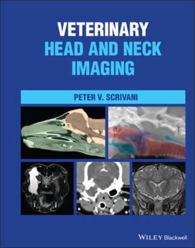
Veterinary Head and Neck Imaging
Peter V. Scrivani
ISBN: 9781119118596
December 2021
Wiley-Blackwell
800 Pages
Description
VETERINARY HEAD AND NECK IMAGING
A complete, all-in-one resource for head and neck imaging in dogs, cats, and horses
Veterinary Head and Neck Imaging is a comprehensive reference for the diagnostic imaging of the head and neck in dogs, cats, and horses. The book provides a multimodality, comparative approach to neuromusculoskeletal, splanchnic, and sense organ imaging. It thoroughly covers the underlying morphology of the head and neck and offers an integrated approach to understanding image interpretation.
Each chapter covers a different area and discusses developmental anatomy, gross anatomy, and imaging anatomy, as well as the physical limitations of different modalities and functional imaging. Commonly encountered diseases are covered at length.
Veterinary Head and Neck Imaging includes all relevant information from each modality and discusses multi-modality approaches. The book also includes:
- A thorough introduction to the principles of veterinary head and neck imaging, including imaging technology, interpretation principles, and the anatomic organization of the head and neck
- Comprehensive explorations of musculoskeletal system and intervertebral disk imaging, including discussions of degenerative diseases, inflammation, and diskospondylitis
- Practical discussions of brain, spinal cord, and cerebrospinal fluid and meninges imaging, including discussions of trauma, vascular, and neoplastic diseases
- In-depth treatments of peripheral nerve, arterial, venous and lymphatic, respiratory, and digestive system imaging
Veterinary Head and Neck Imagingis a must-have resource for veterinary imaging specialists and veterinary neurologists, as well as for general veterinary practitioners with a particular interest in head and neck imaging.
목차
Table of contents
Preface
SECTION 1 INTRODUCTION TO HEAD AND NECK IMAGING IN ANIMALS
1 Some Basic Concepts about Head and Neck Anatomy
1.1 Terms of Location, Orientation, and Movement
1.2 External Features of the Head and Neck
1.3 Overview of Neuroanatomic Localization during Neuroimaging
1.3.1 Divisions of the Central Nervous System
1.3.2 Neuroaxis Localization
1.3.3 Clinical Descriptors for the Location of Intracranial Abnormalities
References
2 Some Basic Concepts about Medical Imaging
2.1 Introduction
2.1.1 What is an Image?
2.1.2 What is Medical Imaging?
2.2 Medical Imaging Devices
2.2.1 Imaging Technologies
2.2.2 Imaging Techniques, Applications, and Examinations
2.3 The Medical Image
2.3.1 Picture Elements and Volumetric Picture Elements
2.3.2 Representing Tissue Characteristics through the Grayscale
2.3.3 Resolution
2.4 Image Evaluation
2.4.1 Getting Started
2.4.2 Imaging Signs and Patterns
2.4.3 Image Evaluation
References
SECTION 2 MUSCULOSKELETAL IMAGING
3 The Musculoskeletal System
3.1 Imaging Anatomy
3.1.1 Bone
3.1.2 Joints and Ligaments
3.1.3 Muscles and Tendons
3.1.3.1 Fascia and Fascial Compartments
3.2 Musculoskeletal Abnormalities
3.2.1 Developmental Malformations
3.2.1.1 Cranium, Face, and Craniocervical Junction
3.2.1.2 Vertebrae
3.2.2. Degenerative Diseases
3.2.2.1 Joints
3.2.2.2 Vertebrae
3.2.3 Inflammatory Diseases
3.2.3.1 Infectious
3.2.3.2 Non-infectious
3.2.4 Neoplasia
3.2.5 Nutritional, Metabolic, Toxic Diseases
3.2.6 Trauma
3.2.6.1 Soft-tissue Trauma
3.2.6.2 Fracture
3.2.6.3 Dislocation
References
4 Intervertebral Disks
4.1 Imaging Anatomy
4.2 Intervertebral Disk Abnormalities
4.2.1 Developmental Malformations
4.2.2 Infection/Inflammation
4.2.3 Trauma
4.2.4 Degeneration
4.2.5 Herniation
References
SECTION 3 NERVOUS SYSTEM IMAGING
5 Cerebrospinal Fluid
5.1 Imaging Anatomy
5.2 CSF Production, Absorption, and Flow
5. 3 Cerebrospinal Fluid Abnormalities
5.3.1 Intra-axial Fluid Accumulations
5.3.2 Extra-axial Fluid Accumulations
5.3.3 Intramedullary Fluid Accumulations
5.2.4 Extramedullary Fluid Accumulations
References
6 The Central Nervous System
6.1 Imaging Anatomy
6.2 Brain and Spinal Cord Abnormalities
6.2.1 Imaging Patterns of CNS Disease
6.2.1.1 Some Additional Imaging Signs
6.2.1.2 Contrast Enhancement
6.2.2. Secondary Intracranial Abnormalities
6.2.2.1 Intracranial Hypertension
6.2.2.2 Cerebral Edema
6.2.2.3 MRI Signs Induced by Seizures
6.2.2.4 Brain Herniation
6.2.2 Developmental Malformations
6.2.3 Vascular Disorders
6.2.3.1 Ischemia
6.2.3.2 Hemorrhage
6.2.3.3 Hemorrhagic Infarction
6.2.4 Trauma
6.2.4.1 Traumatic Brain Injury
6.2.4.2 Traumatic Spinal Cord Injury
6.2.5 Neoplasia
6.2.6 Inflammatory Diseases
6.2.6.1 Infectious
6.2.6.2 Non-Infectious
6.2.7 Degenerative Diseases
References
7 The Peripheral Nervous System
7.1 Imaging Anatomy
7.1.1 Cranial Nerves
7.1.2 Spinal Nerves
7.1.2.1 The Cervical Nerves
7.1.2.2 The Brachial Plexus
7.1.2.3 The Sympathetic Division
7.2 Peripheral Nerve Abnormalities
7.2.1 Neoplasia
7.2.2 Trauma
7.2.3 Inflammatory Diseases
7.2.4 Compression
7.2.1 Degenerative Diseases
References
SECTION 4 SPLANCHNIC (VISCERA), VASCULAR, AND SENSE ORGAN IMAGING
8 The Digestive System
8.1 Imaging Anatomy
8.1.1 Oral Cavity
8.1.2 Pharynx
8.1.3 Cervical Esophagus
8.2 Digestive Track Abnormalities
8.2.1 Developmental Malformations
8.2.2 Dysphagia
8.2.3 Neoplasia
8.2.4 Inflammation
References
9 The Respiratory System
9.1 Imaging Anatomy
9.1.1 Nasal Cavities and External Nose
9.1.2 Paranasal Sinuses
9.1.3 Nasopharynx, Larynx, and Cervical Trachea
9.2 Respiratory Track Abnormalities
9.2.1. Developmental Anomalies
9.2.2. Inflammation/Infection
9.2.3. Neoplasms
9.2.4. Degenerative Disorders
References
10 Sense Organs, Circulatory System, and Endocrine System
10.1 Imaging Anatomy
10.1.1 Eye
10.1.2 Ear
10.1.3 Circulatory System
10.1.4 Endocrine System
10.2 Orbital Disorders
10.2.1 Trauma
10.2.2 Inflammatory Disease
10.2.3 Neoplasms
10.3 Ear Disorders
10.3.1 Ear Diseases
10.3.2 Guttural Pouch Disease
10.3.3 Imaging Patterns of Disease
10.4 Circulatory and Endocrine Disorders
10.4.1 Developmental Anomalies
10.4.2 Endocrine Disorders
10.4.3 Circulatory System Disorders
References
Index
About the Author
배송 정보
- 배송 방법 : 택배
- 배송 지역 : 전국지역
- 배송 비용 : 3,000원
- 배송 기간 : 2일 ~ 5일
- 배송 안내 : - 산간벽지나 도서지방은 별도의 추가금액을 지불하셔야 하는 경우가 있습니다.
고객님께서 주문하신 상품은 입금 확인후 배송해 드립니다. 다만, 상품종류에 따라서 상품의 배송이 다소 지연될 수 있습니다.
고객님께서 주문하신 도서라도 도매상 및 출판사 사정에 따라 품절/절판 등의 사유로 취소될 수 있습니다. 입금확인이 되면 익일발송(주말,공휴일제외)을 원칙으로 합니다.
교환 및 반품 정보
교환 및 반품이 가능한 경우
- 상품을 공급 받으신 날로부터 7일이내 내용이 파본일 경우
단, 비닐포장을 개봉하였거나 내용물이 훼손되어 상품가치가 상실된 경우에는 교환/반품이 불가능합니다.
- 공급받으신 상품 및 용역의 내용이 표시.광고 내용과
다르거나 다르게 이행된 경우에는 공급받은 날로부터 3월이내, 그사실을 알게 된 날로부터 30일이내
교환 및 반품이 불가능한 경우
- 고객님의 책임 있는 사유로 상품등이 멸실 또는 훼손된 경우. 단, 상품의 내용을 확인하기 위하여
포장 등을 훼손한 경우는 제외
- 포장을 개봉하였거나 포장이 훼손되어 상품가치가 상실된 경우
- 고객님의 사용 또는 일부 소비에 의하여 상품의 가치가 현저히 감소한 경우
- 시간의 경과에 의하여 재판매가 곤란할 정도로 상품등의 가치가 현저히 감소한 경우
- 복제가 가능한 상품등의 포장을 훼손한 경우
(자세한 내용은 고객만족센터 1:1 E-MAIL상담을 이용해 주시기 바랍니다.)
※ 고객님의 마음이 바뀌어 교환, 반품을 하실 경우 상품반송 비용은 고객님께서 부담하셔야 합니다.


